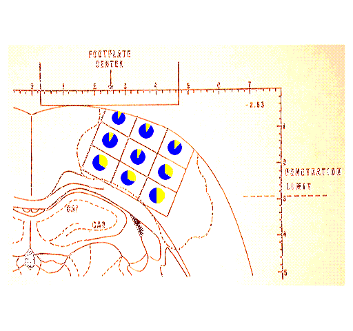
| Figure 1. Drawing of a section of rat brain at the stereotaxic plane through the middle of the impact site. The footplate resting on the dura is struck by a 20 g weight falling 20 cm (described as 400 g/cm as the area of the footplate is one cm.). The dashed line drawn in the cortex indicates the area of a fully developed cyst overlapping in 6 animals examined at 25-30 days after injury. Within, this area is the sampling grid. that was superimposed upon sections taken from sections from rats sacrificed early after injury prior to the development of the cavity. The pie charts within each sector indicate the approximate proportion of argyrophilic neurons at 24 hours after injury. Note the marked sparing of neurons in the superficial laminae and the greater cell death in the deep lateral sector. Similar results were seen using thionine and acid fucshin studies. |