![]()
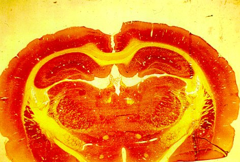
Ablation illustration from:
Sutton, RL, Hovda, DA, Chen. MJ & Feeney, DM. Alleviation of Brain Injury-Induced Cerebral Metabolic Depression by Amphetamine: A Cytochrome Oxidase Histochemistry Study. Neural Plasticity In Press, 1998.
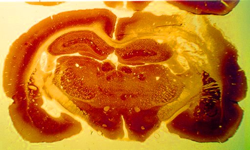
TBI illustration from:
Weisend, M.P., R.A. Salazar & D.M. Feeney. Sensorimotor contusion but not ablation increases cytochrome oxidase activity in rat auditory and entorhinal cortex. (1990) Proc. 8th Ann. Mtg.. Neurotrauma Society, St. Louis, MO.
For a general review of the Cytochrome Oxidase Histochemistry method, see:
Wong-Riley, M.T.T. (1989) Cytochrome oxidase: an endogenous metabolic marker for neuronal activity. TINS, 12, 94-101.
For details regarding the methods see:
Wong-Riley MTT (1979) Changes in the visual system of monocularly sutured or enucleated cats demonstrable with cytochrome oxidase histochemistry. Brain Res 171:11-28.
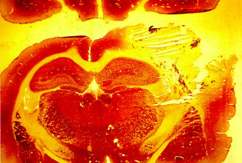
Densitometry measures indicate that compared to sham operate controls (labeled "Normal", ablation of the right hemisphere sensorimotor cortex produces a reduction of Cytochrome Oxidase (CYO) levels at 2 days after surgery. This is significant bilaterally in cerebral cortex, red nucleus and superior Colliculi and in the ipsilateral nucleus accumbens, caudate-putamen and globus pallidus. In contrast, after weight-drop impact trauma, in addition to paling in neocortex there is a marked increase in CYO in the ipsilateral temporal lobe. This is not a stain artifact since there is an increase in regional cerebral blood flow in the same area after trauma. See Figure 3 on this web site.
It is more likely that the increase in CYO mitochondrial enzyme and accompanying increase in rCBF results from epileptiform activity observed in this TBI model and reported in the following references.
Nilsson P, Ronne-Engstrom E, Flink R, Ungerstedt U, Carlson H Hillered L (1994) Epileptic seizure activity in the acute phase following cortical impact trauma in rat. Brain Res 637:227-232.
Krobert KA, Salazar RA, Sutton RL, Feeney DM (1992) Temporal evolution of histopathology and unit activity in rat hippocampal CA3 region after focal cortical contusion. J Neurotrauma 9:64.
Golarai, D. M. Feeney & J. A. Connor Epileptiform Responses in Cortical Slices After Traumatic Brain Injury. Neuroscience Abstracts, 25, 1999. In Press.
![]()