Click on image or hypertext for a larger view.
|
The Nervous System - Spinal Cord and Peripheral Nerves | |

|
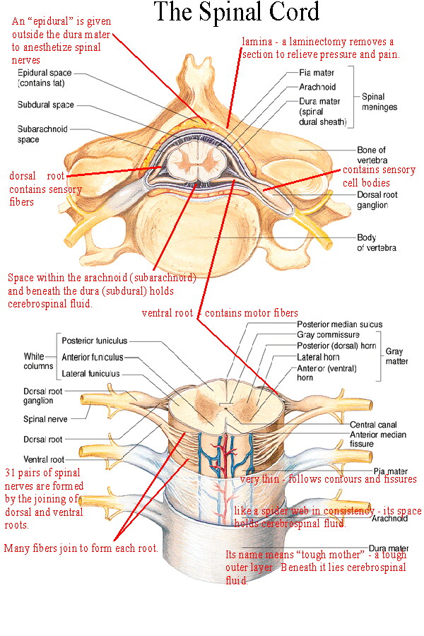
The spinal cord is the connection center for the reflexes as well as the afferent (sensory) and efferent (motor) pathways for most of the body below the head and neck. The spinal cord begins at the brainstem and ends at about the second lumbar vertebra. The sensory, motor, and interneurons discussed previously are found in specific parts of the spinal cord and nearby structures. Sensory neurons have their cell bodies in the spinal (dorsal root) ganglion. Their axons travel through the dorsal root into the gray matter of the cord. Within the gray matter are interneurons with which the sensory neurons may connect. Also located in the gray matter are the motor neurons whose axons travel out of the cord through the ventral root. The white matter surrounds the gray matter. It contains the spinal tracts which ascend and descend the spinal cord. Surrounding both the spinal cord and the brain are the meninges, a three layered covering of connective tissue. The dura mater is the tough outer layer. Beneath the dura is the arachnoid which is like a spider web in consistency. The arachnoid has abundant space within and beneath it (the subarachnoid space) which contains cerebrospinal fluid, as does the space beneath the dura mater (subdural space). This cerebrospinal fluid supplies buoyancy for the spinal cord and brain to help provide shock absorption. The pia mater is a very thin layer which adheres tightly to the surface of the brain and spinal cord. It follows all contours and fissures (sulci) of the brain and cord. |
| Terms:
ganglion - a collection of cell bodies located outside the Central Nervous System. The spinal ganglia or dorsal root ganglia contain the cell bodies of sensory neurons entering the cord at that region. nerve - a group of fibers (axons) outside the CNS. The spinal nerves contain the fibers of the sensory and motor neurons. A nerve does not contain cell bodies. They are located in the ganglion (sensory) or in the gray matter (motor). tract - a group of fibers inside the CNS. The spinal tracts carry information up or down the spinal cord, to or from the brain. Tracts within the brain carry information from one place to another within the brain. Tracts are always part of white matter. gray matter - an area of unmyelinated neurons where cell bodies and synapses occur. In the spinal cord the synapses between sensory and motor and interneurons occurs in the gray matter. The cell bodies of the interneurons and motor neurons also are found in the gray matter. white matter - an area of myelinated fiber tracts. Myelination in the CNS differs from that in nerves. | |
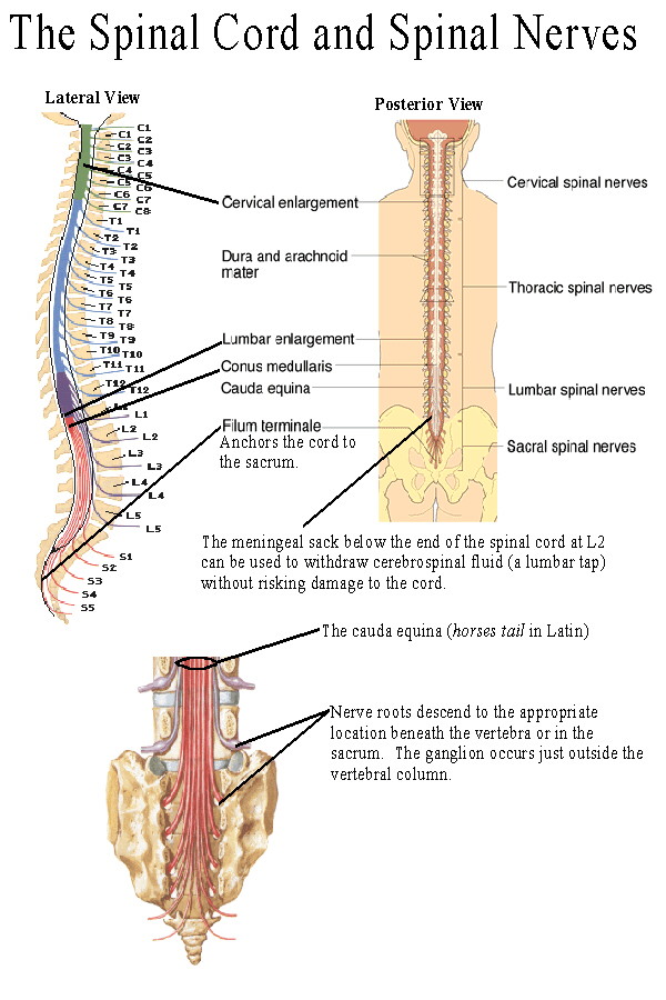 |
At 31 places along the spinal cord the dorsal and ventral roots come together to form spinal nerves. Spinal nerves contain both sensory and motor fibers, as do most nerves. Spinal nerves are given numbers which indicate the portion of the vertebral column in which they arise. There are 8 cervical (C1-C8), 12 thoracics (T1-T12), 5 lumbar (L1-L5), 5 sacral (S1-S5), and 1 coccygeal nerve. Nerve C1 arises between the cranium and atlas (1st cervical vertebra) and C8 arises between the 7th cervical and 1st thoracic vertebra. All the others arise below the respective vertebra or former vertebra in the case of the sacrum. Since the actual cord ends at the second lumbar vertebra, the later roots arise close together on the cord and travel downward to exit at the appropriate point. These nerve roots are called the cauda equina because of their resemblance to a horses tail. |
| The dermatomes are somatic or musculocutaneous areas served by fibers from specific spinal nerves. The map of the dermatomes is shown by Figure 13.11.This map is useful in diagnosing the origin of certain somatic pain, numbness, tingling etc. when these symptoms are caused by pressure or inflammation of the spinal cord or nerve roots. Referred pain is caused when the sensory fibers from an internal organ enter the spinal cord in the same root as fibers from a dermatome. The brain is poor at interpreting visceral pain and instead interprets it as pain from the somatic area of the dermatome. So pain in the heart is often interpreted as pain in the left arm or shoulder, pain in the diaphragm is interpreted as along the left clavicle and neck, and the "stitch in your side" you sometimes feel when running is pain in the liver as its vessels vasoconstrict. (See Figure 14.8) | |
| Spinal nerves join together in plexuses. (See Figure 13.5) A plexus is an interconnection
of fibers which form new combinations as the "named" or peripheral nerves. There are
four voluntary plexuses (there are some autonomic plexuses which will be mentioned
later): they are the cervical plexus, the brachial plexus, the lumbar plexus, and the
sacral plexus. Each plexus gives rise to new combinations of fibers as the peripheral
nerves. The nerves and plexuses you need to know are:
Cervical Plexus (See Figure 13.7, Table 13.3) - the phrenic nerve travels through the thorax to innervate the diaphragm. Brachial Plexus (See Figure 13.8, Table 13.4) -
Lumbar Plexus (See Figure 13.9, Table 13.5)
Sacral Plexus (See Figure 13.10, Table 13.6)
| |
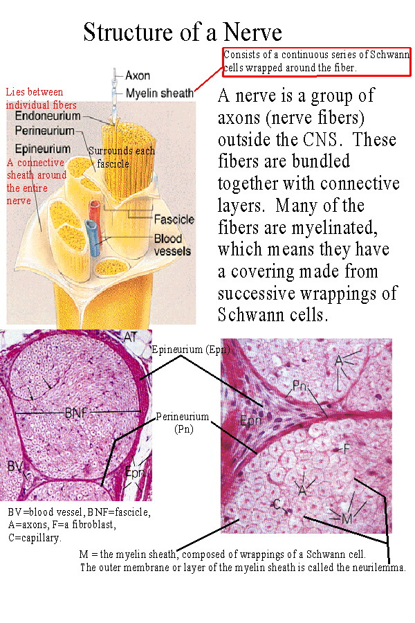 |
Structure of a nerve:
A peripheral nerve is arranged much like a muscle in terms of its connective tissue. It has an outer covering which forms a sheath around the nerve, called the epineurium. Often a nerve will run together with an artery and vein and their connective coverings will merge. Nerve fibers, which are axons, organize into bundles known as fascicles with each fascicle surrounded by the perineurium. Between individual nerve fibers is an inner layer of endoneurium. |
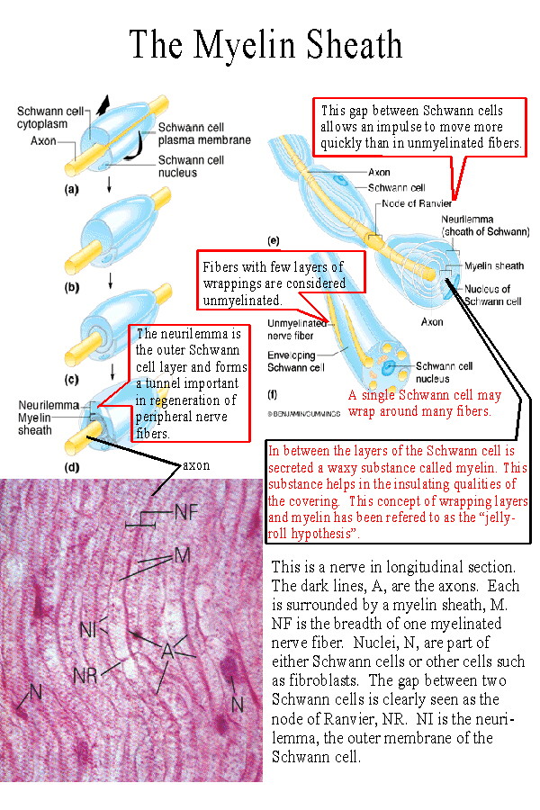 |
The myelin sheath in peripheral nerves consists of Schwann cells
wrapped in many layers around the axon fibers. Not all fibers in a
nerve will be myelinated, but most of the voluntary fibers are. The
Schwann cells are portrayed as arranged along the axon like
sausages on a string. (A more apt analogy would be like jelly rolls!)
Gaps between the Schwann cells are called nodes of Ranvier.
These nodes permit an impulse to travel faster because it doesn't
need to depolarize each area of a membrane, just the nodes. This
type of conduction is called saltatory conduction and means that
impulses will travel faster in myelinated fibers than in unmyelinated
ones.
The myelin sheath does several things: 1) It provides insulation to help prevent short circuiting between fibers. Diseases which destroy the myelin sheath lead to inability to control muscles, perceive stimuli etc. One such disease is multiple sclerosis, an autoimmune disorder in which your own lymphocytes attack the myelin proteins. [See Beta Interferon and Multiple Sclerosis]. 2) The myelin sheath provides for faster conduction. 3) The myelin sheath provides for the possibility of repair of peripheral nerve fibers. Schwann cells help to maintain the micro-environments of the axons and their tunnel (the neurilemma tunnel) permits re-connection with an effector or receptor. (See below) CNS fibers, not having the same type of myelination accumulate scar tissue after damage, which prevents regeneration. [See Spinal Cord Repair] |
| Regeneration of a peripheral nerve fiber (See Figure 13.3) depends upon several things. First the damage must be far from the cell body. Anterograde degeneration destroys the axon distal to the point of damage. Retrograde degeneration causes the fiber to degenerate for a distance back toward the cell body. The amount of axoplasm lost determines whether the neuron can survive. Secondly the myelin sheath and its neurilemma tunnel must be intact. Chemicals such as the myelin proteins tend to inhibit regrowth, but macrophages will enter the damaged area and phagocytize these proteins and other debris. Schwann cells will proliferate and secrete growth stimulating factors and provide the chemical and physical needs necessary for growth and re-innervation by the axon. | |
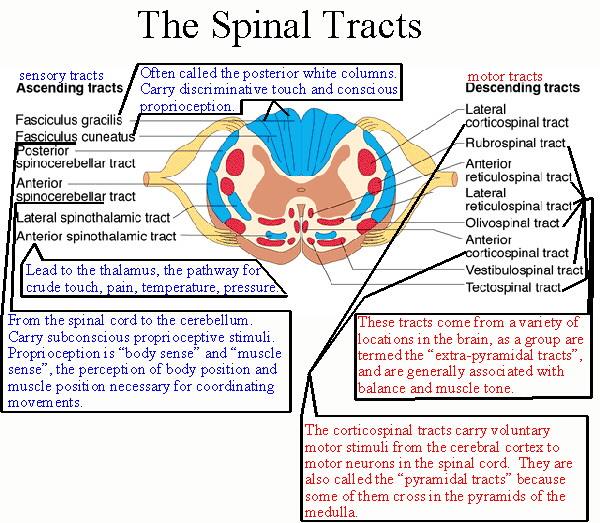 |
The Spinal Tracts: (See Figure 12.30)
The white matter of the spinal cord contains tracts which travel up and down the cord. Many of these tracts travel to and from the brain to provide sensory input to the brain, or bring motor stimuli from the brain to control effectors. Ascending tracts, those which travel toward the brain are sensory, descending tracts are motor. Figure 12.30 shows the location of the major tracts in the spinal cord. For most the name will indicate if it is a motor or sensory tract. Most sensory tracts names begin with spino, indicating origin in the spinal cord, and their name will end with the part of the brain where the tract leads. For example the spinothalamic tract travels from the spinal cord to the thalamus. Tracts whose names begin with a part of the brain are motor. For example the corticospinal tract begins with fibers leaving the cerebral cortex and travels down toward motor neurons in the cord. [See Spinal Tract Pathways] |
NEXT: The Reflexes