The
Myograph
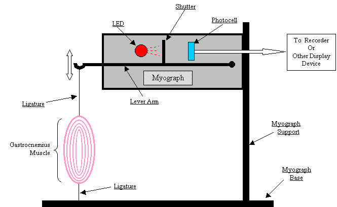
The myograph (lit. ‘muscle writer’) is a device we use to measure the force generated by a contracting muscle. Myographs are examples of transducers that convert force into an electrical output that can be displayed on an oscilloscope or a computer monitor. The relevant components of a myograph are:
1. a light source, either a light bulb or a light emitting diode (LED);
2. a photocell;
3. a lever arm, which is connected to
4. a shutter.
A schematic diagram of a typical myograph as it would be set up to measure tension generated by the gastrocnemius muscle would be:

Light reaching the photocell from the lamp will cause it to emit a current that can be detected and displayed by some sort of recording device (in our case, the display produced by the simulation). As shown in the schematic, when there is no tension applied to the lever, the shutter completely prevents any light from reaching the photocell, and there will be no current produced. Correspondingly, a reading of zero will be displayed by the recorder.
When tension is applied to the lever, for example by contractile activity in the gastrocnemius muscle, the lever will deflect downward, pulling the shutter down and deshielding the photocell. Since the current output from the photocell is proportional to the photon flux (i.e., the more photons striking the photocell, the more current is produced), we have a good way to monitor the amount of deflection of the lever. And, since the deflection is pretty much a linear function of the force ( = tension) being applied to the lever, we have a fairly precise measure of the tension.
You will use the myograph, or something similar, in a number of the simulations you will be conducting throughout the semester, so you’ll want to familiarize yourself with how it works.