| This is a Visual of the heart going through a cardiac cycle with an EKG depiction (of what it looks like on the EKG) as well as a ventricular myocyte going through the Phases of the Depolarization/Repolarization timeline. |
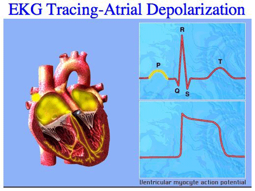 |
| During Atrial Depolarization, the ventricular myocytes are in Phase 4 of the Depolarization/Repolarization timeline. The ventriclar myocytes are restoring ions with the Na+/K+ Pump. |
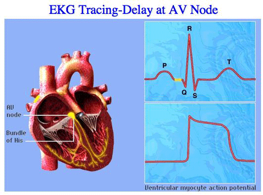 |
| This delay at the AV Node gives the heart a little more time to pump all of the blood in the atria into the ventricles. |
|
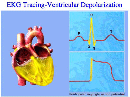 |
| The ventricle myocyte is going through Phase 0: Depolarization. There is an influx of Na+ via the FAST Na+ channels. |
|
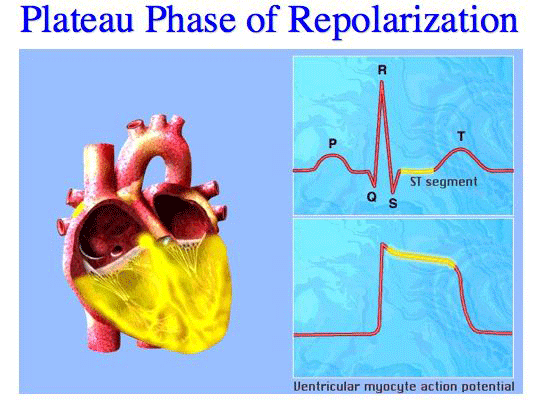 |
| Please note that Phase 1, the Early Rapid Repolariation is so quick it is not seen on the EKG. In Phase 1, the FAST Na+ channels close and K+ begins to efflux. SHOWN in the figure above is Phase 2 (Slow Repolarization, the Plateau phase) where there is an efflux of K+ and influx of Ca++ and Na+ (via the SLOW Na+ channels). |
|
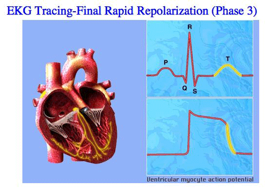 |
| In Phase 3, Final Rapid Repolarization, K+ continues to efflux and the Ca++ and SLOW Na+ channels close. |


