
Click on diagrams and hypertext for bigger views. Click on ![]() to get specific histology images.
to get specific histology images.
|
Muscular Histology & Structure | |
| There are three types of muscle tissue, all of which share some common properties:
excitability or responsiveness - muscle tissue can be stimulated by electrical, physical, or chemical means. contractility - the response of muscle tissue to stimulation is contraction, or shortening. elasticity or recoil - muscles have elastic elements (later we will call these their series elastic elements) which cause them to recoil to their original size. stretchability or extensibility - muscles can also stretch and extend to a longer-than-resting length. | |
 |
The three types of muscle: skeletal, cardiac, and visceral (smooth)
muscle. [See Muscle Histology]
You should make a chart similar to Table 9.4 and the one from class.
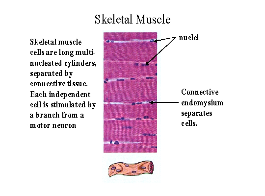
Skeletal muscle is found attached to the bones for movement. Its cells are long multi-nucleated cylinders. They acquired this characteristic because they develop from the fusion of small single cells into long units. The cells may be many inches long but vary in diameter, averaging between 100 and 150 microns. Skeletal muscle cells are independent cells separated from one another by connective tissue and must each be stimulated by axons of a neuron. All the cells innervated by branches from the same neuron will contract at the same time and are referred to as a motor unit. Motor units vary in size: large motor units with more than 100 cells are typical of the slow acting postural muscles. Very small motor units with around 10 cells or so are typical of fast acting muscles with very precise control such as those which move the eye. Most of our muscles have a mixture of motor units of different sizes. Skeletal muscle is voluntary because the neurons which innervate it come from the somatic or voluntary branch of the nervous system. That means you have willful control over your skeletal muscles. Skeletal muscles have distinct stripes or striations which identify them and are related to the organization of protein myofilaments inside the cell. |
| Cardiac muscle is the muscle found in the heart. It is composed of much shorter cells than skeletal muscle which branch to connect to one another. These connections are by means of gap junctions called intercalated disks which allow an electrochemical impulse to pass to all the connected cells. This causes the cells to form a functional network called a syncytium in which the cells work as a unit. Faint striations indicate a similar, but not identical, arrangement of myofilaments in cardiac and skeletal muscle. Many cardiac muscle cells are myogenic which means that the impulse arises from the muscle, not from the nervous system. This causes the heart muscle and the heart itself to beat with its own natural rhythm. But the autonomic nervous system controls the rate of the heart and allows it to respond to stress and other demands. As such the heart is said to be involuntary. | |
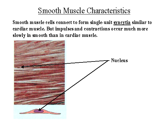
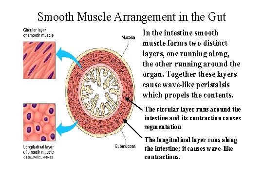
| Visceral muscle is found in the body's internal organs and blood vessels. It is usually called smooth muscle because it has no striations and is therefore smooth in appearance. It is found as layers in the mucous membranes of the respiratory and digestive systems. It is found as distinct bands in the walls of blood vessels and as sphincter muscles. Single unit smooth muscle is also connected into a syncytium similar to cardiac muscle and is also partly myogenic. As such it causes continual rhythmic contractions in the stomach and intestine. There and in blood vessels smooth muscle also forms multiunit muscle which is stimulated by the autonomic nervous system. So smooth muscle is involuntary as well. See also Figure 9.21 (modified). | |
 |
Structure and function of skeletal muscle.
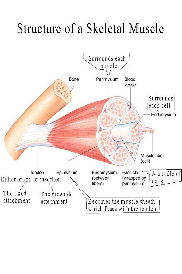
Skeletal muscles have a belly which contains the cells and which attaches by means of tendons or aponeuroses to a bone or other tissue. An aponeurosis is a broad, flat, tendinous attachment, usually along the edge of a muscle. A muscle attaches to an origin and an insertion. The origin is the more fixed attachment, the insertion is the more movable attachment. A muscle acts to shorten, pulling the insertion toward the origin. A muscle can only pull, it cannot push. Muscles usually come in pairs of antagonistic muscles. The muscle performing the prime movement is the agonist, the opposite acting muscle is the antagonist. When the movement reverses, the names reverse. For example, in flexing the elbow the biceps brachii is the agonist, the triceps brachii is the antagonist. When the movement changes to extension of the elbow, the triceps becomes the agonist and the biceps the antagonist. Antagonists are never totally relaxed. Its function is to provide control and damping of movement by maintaining tone against the agonist. This is called eccentric movement. Muscles can also act as synergists, working together to perform a movement. This movement can be different from that performed when the muscles work independently. For example, the sternocleidomastoid muscles each rotate the head in a different direction. But as synergists they flex the neck. Fixators act to keep a part from moving. For example fixators act as postural muscles to keep the spine erect and the leg and vertebral column extended when standing. Fixators such as the rhomboids and levator scapulae keep the scapula from moving during actions such as lifting with the arms. Your assignment is to make a chart of the muscles showing their location and action. Look for examples of antagonists, synergists, and fixators. You may find the Muscle Action Page helpful in identifying these characteristics. |