
Click on images or hypertext for a bigger view. Click on ![]() for histology
module.
for histology
module.
Links to ![]()
|
The Nervous System - Neurology | |
| Functions of the nervous system:
1) Control of effectors, both voluntary and autonomic - muscles (skeletal, smooth, cardiac), glands. 2) Response to stimuli 3) Integration of processes 4) Responsible for conscious thought and perception, emotions, personality, the mind.
Structural Divisions of the nervous system: 1) Central Nervous System (CNS) - the brain and spinal cord. 2) Peripheral Nervous System (PNS) - the nerves, ganglia, receptors, etc.
Functional Divisions of the Nervous System: 1) The Voluntary Nervous System - (a.k.a. somatic division) control of willful control of effectors (skeletal muscles) and conscious perception. Mediates voluntary reflexes. 2) The Autonomic Nervous System - control of autonomic effectors - smooth muscles, cardiac muscle, glands. Responsible for "visceral" reflexes. | |
| Nervous System Histology - cell types: (See Figure 11.3)
1) Neurons - the functional cells of the nervous system. See below. 2) Neuroglia (glial cells) - Long described as supporting cells of the nervous system, there is also a functional interdependence of neuroglial cells and neurons. [See Glioma Tumors] a) astrocytes - these cells anchor neurons to blood vessels, regulate the micro-environment of neurons, and regulate transport of nutrients and wastes to and from neurons. [See Blood-Brain Barrier] b) microglia - these cells are phagocytic to defend against pathogens. They may also monitor the condition of neurons. c) ependymal cells - these cells line the fluid-filled cavities of the brain and spinal cord. They play a role in production, transport, and circulation of the cerebrospinal fluid. d) oligodendrocyte - produce the myelin sheath in the CNS which insulates and protects axons as well as providing for saltatory conduction. e) Schwann cells - produce the myelin sheath in the PNS. The myelin sheath protects and insulates axons, maintains their micro-environment, and enables them to regenerate and re-establish connection with receptors or effectors. Enables saltatory conduction. f) satellite cells - surround cell bodies of neurons in ganglia. Their role is to maintain the micro-environment and provide insulation for the ganglion cells. | |
 |
Neuron Structure:
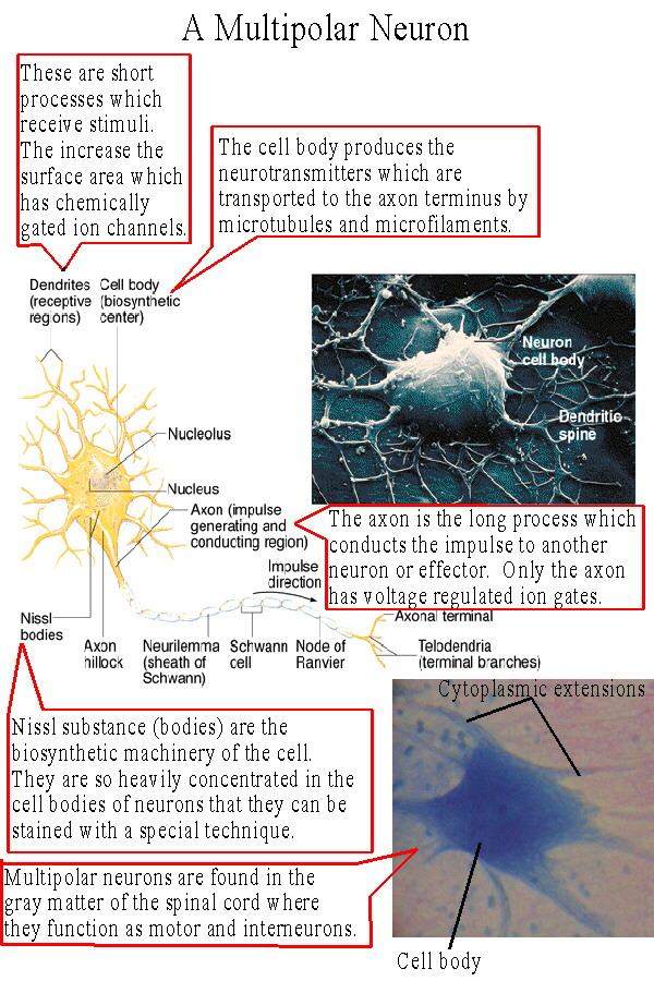
In order to connect to other cells, receptors, and effectors, neurons have cytoplasmic extensions which attach to an enlarged area known as the cell body or cyton. Within the cell body is the nucleus and the neuron's biosynthetic machinery, the rough endoplasmic reticulum and the Golgi bodies. These organelles are so highly concentrated they can be visualized with a light microscope when stained with a specific technique. Called Nissl substance after the scientist who invented the staining technique, they manufacture the neurotransmitters which the neuron must secrete in large quantities. The neurotransmitter molecules are transported to the axon terminus by microfilaments and microtubules. There are two basic types of cytoplasmic extensions: the dendrites and the axon. Dendrites are short branching processes which receive stimuli from receptors or other neurons. They can perform this function because they, like the exposed membrane of the cell body, possess chemically regulated ion gates (chemically gated ion channels) which respond to stimulation by neurotransmitters. So the dendrites increase the area on which a neuron can be stimulated and together with the rest of the membrane of the cell body constitute the neuron's receptive region (See Table 11.1). A neuron will usually have only one axon, although it may branch extensively. The axon has voltage regulated ion gates (voltage gated ion channels) and therefor is responsible for carrying an impulse to another neuron or effector. The axon represents the neuron's conducting region. At the end of the axon, the axon terminus, is the secretory region where the neurotransmitters are released into the synapse. The trigger zone is where the area with chemically regulated gates and the area with voltage regulated gates meet, usually at the junction of the axon and cell body, the axon hillock. In this area summation of depolarization and hyperpolarization (explained later) can produce enough depolarization to open the voltage regulated gates and produce an action potential. |
 |
Types of neurons based on structure:
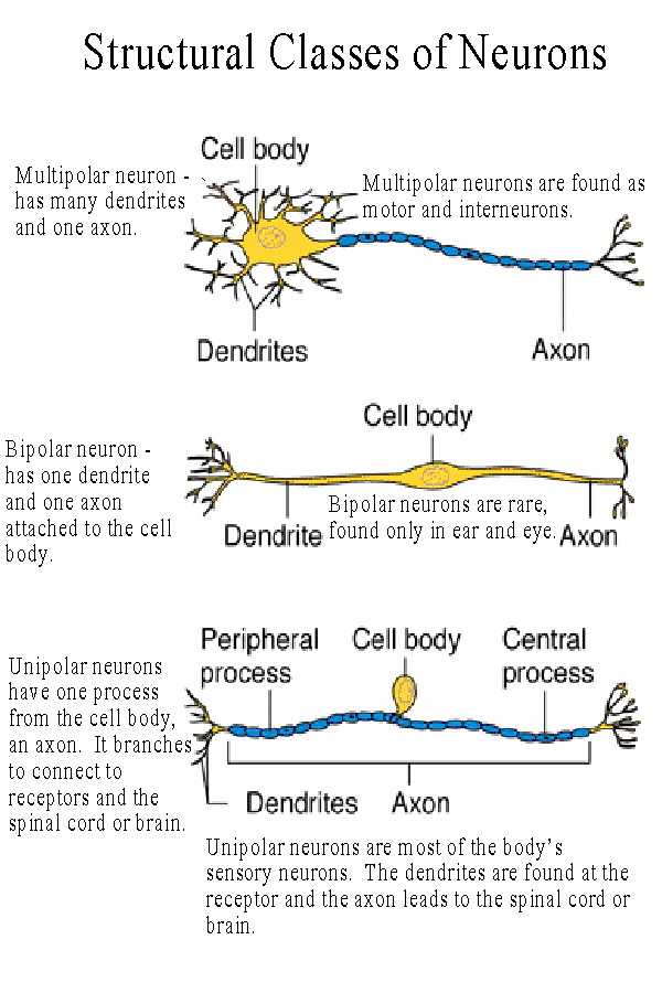
The neuron pictured in Figure 11.4 and linked above is a multipolar neuron because it has many poles or processes, the dendrites and the axon. Multipolar neurons are found as motor neurons and interneurons(see below). There are also bipolar neurons with two processes, a dendrite and an axon, and unipolar neurons, which have only one process, classified as an axon. (See Table 11.1). Unipolar neurons are found as most of the body's sensory neurons. Their dendrites are the exposed branches connected to receptors, the axon carries the action potential in to the central nervous system. |
 |
Types of neurons based on function:
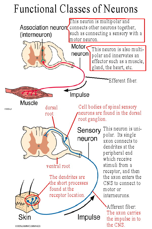
motor neurons - these carry a message to a muscle, gland, or other effector. They are said to be efferent, i.e. they carry the message away from the central nervous system. sensory neurons - these carry a message in to the CNS. They are afferent, i.e. going toward the brain or spinal cord. interneuron (a.k.a. association neuron, connecting neuron) - these neurons connect one neuron with another. For example in many reflexes interneurons connect the sensory neurons with the motor neurons. |
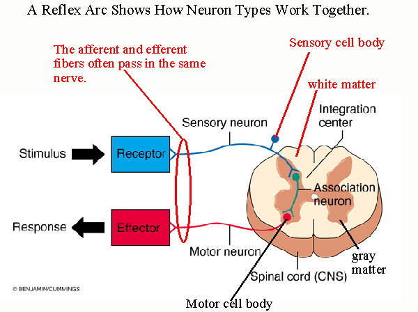 |
A simple Reflex Arc illustrates how these three types of neurons might work together. We will discuss types of reflexes in a later section. The reflex shown has its center in the spinal cord our next topic. |